Paralyzed Men Gain Movement Without Surgery
« A noninvasive treatment helped 5 men with complete muscle paralysis in the lower body voluntarily move their legs in a step-like pattern.
The finding suggests that stimulation may help reactivate dormant nerve connections between the brain and spinal cord in some paralyzed patients.
The spinal cord is the central nerve cable connecting the brain to the rest of the body. Damaged can lead to serious disabilities, including paralysis. More than a quarter of a million Americans are now living with spinal cord injuries.
Last year, NIH-funded researchers reported that a surgically implanted stimulating device allowed 4 men to regain some leg movement after spinal cord injuries had left their voluntary muscles completely paralyzed below the chest.
In a follow-up study, the scientists—led by Drs. V. Reggie Edgerton and Yury Gerasimenko of the University of California, Los Angeles—tested a nonsurgical strategy for stimulating the spinal cord. Called transcutafneous stimulation, the method delivers electrical current to the spinal cord via electrodes strategically placed on the skin over the spine.
The work was funded in part by NIH’s National Institute of Biomedical Imaging and Bioengineering (NIBIB), National Center for Advancing Translational Sciences (NCATS), and other NIH components. Results were reported in the online edition of Journal of Neurotrauma.
Five men—each paralyzed for more than 2 years—received 18 weekly sessions of the spinal stimulation for about 45 minutes. The sessions also included muscle conditioning, in which therapists manually moved the patients’ legs in a step-like pattern. During the final 4 weeks, the men received twice-daily doses of buspirone. This drug has been shown to induce mobility in mice with spinal cord injuries.
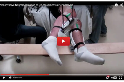
The men were instructed at different points during spinal stimulation to try to move their legs or to remain passive. During these sessions, their legs were supported by braces hung from the ceiling, so they could move without resistance from gravity.
Initially, the men’s legs moved only when spinal stimulation was strong enough to generate involuntary step-like movements. But after 4 weeks, the men were able to double their range of motion when voluntarily moving their legs during stimulation. The addition of buspirone further improved their movements and by the end of the study, they were able to move their legs with no stimulation at all. On average, their range of movement equaled that during spinal stimulation.
The researchers recorded electrical signals of the men’s calf muscles while they attempted to flex their feet during stimulation. Over time, the signals increased with the same amount of stimulation, suggesting a re-establishment of communication between the brain and spinal cord.
“It’s as if we’ve reawakened some networks so that once the individuals learned how to use those networks, they become less dependent and even independent of the stimulation,” Edgerton says.
Although the movements achieved in this study aren’t comparable to walking, the results represent progress toward a potential therapy for spinal cord injury. The team is now assessing whether these 5 men can be trained to fully bear their weight, an accomplishment that the 4 men with surgically implanted stimulators have achieved.
Video shows range of voluntary movement before and after treatment. The subject’s legs are supported so that they can move without resistance from gravity. The electrodes are used to record muscle activity.
Credit: Edgerton laboratory/UCLA. »
http://www.nih.gov/researchmatters/august2015/08102015paralyzed.htm


